The Department of Thoracic Surgery of the Ninth Hospital successfully saved the lungs for patients, with a 37cm giant fibrous tumor almost occupying the entire right chest cavity surgery | Tumor | Thoracic Surgery
51 year old Ms. Zhu was diagnosed with a fibroadenoma with a diameter of about 20 centimeters in the right chest cavity at a local hospital in Fujian a year ago. Although she underwent multiple interventional surgeries such as vascular occlusion and radioactive particle implantation, more than a month ago, the examination showed that the tumor had developed to a maximum diameter of about 29 centimeters, almost occupying the entire right chest cavity. Two weeks ago, at the Ninth People's Hospital affiliated with Shanghai Jiao Tong University School of Medicine, this huge tumor measuring 37 cm x 36 cm x 7.6 cm was completely removed, and the lungs were successfully preserved. After 11 days of surgery, the patient recovered well and was discharged smoothly.
According to Ji Guangyu, the deputy chief physician of the thoracic surgery department, Ms. Zhu had already developed symptoms such as difficulty breathing and was unable to lie flat, so she could only sit halfway. After detailed inquiries about medical history and past surgical conditions, as well as reviewing various examination reports and imaging data, the doctors believe that there is still a chance for surgery. At that time, patients and their families also firmly stated that as long as there was one percent hope, they should actively strive for it.
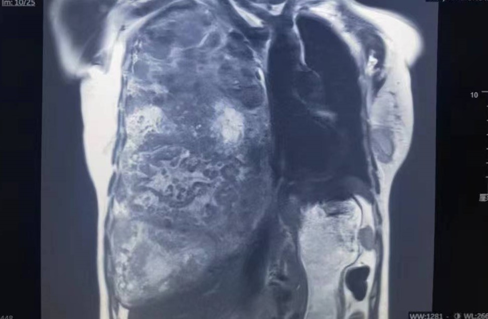
Preoperative magnetic resonance imaging and other examinations indicate a huge thoracic tumor with complete atelectasis in the right lower middle lung. This situation is rare, and if surgery is performed, there are many difficulties: does the tumor invade the pleura and lungs? Can the tumor be completely removed? Does the right lung need to be removed? What is the extent of excision? Is the risk of anesthesia controllable as a huge tumor compresses the mediastinum, causing significant left deviation of the heart and trachea? There may be a lot of intraoperative bleeding, how to ensure the safety of intraoperative blood use?
To this end, the medical department organized multidisciplinary discussions among experts in thoracic surgery, anesthesia, imaging, pathology, transfusion, and critical care medicine. Confirming that the tumor is benign, with intact capsule, and can be operated on, although there is a high risk, it must be overcome.
The surgery was jointly performed by the director of thoracic surgery, Wang Mingsong, and Ji Guangyu. The deputy chief physician, Wang Feng, and attending physician, Wu Jinlong, took the stage together. The tumor was quickly and completely removed within 10 minutes, and the wound was filled with gauze to stop bleeding and the tumor pedicle was stopped bleeding. Further investigation revealed that the tumor pedicle was located on the diaphragm, with a diameter of about 2.5 centimeters. The remaining part of the tumor had intact capsule and almost no adhesion to the pleura and lungs. The right upper lung was dilated, and it was difficult to dilate the middle and lower lungs. It was decided not to remove the tumor immediately, and the surgery was successfully completed within 2 hours. On the 5th day after surgery, the patient moved to the ground and underwent a chest CT scan on the 10th day. Most of the right middle and lower lungs were dilated. After removing all thoracic drainage tubes, the patient was discharged smoothly.
Ji Guangyu explained that Ms. Zhu's preoperative pathology confirmed that it was a solitary fibrous tumor in the chest cavity. Most of these tumors were benign, usually with a capsule, and some were malignant. Preoperative magnetic resonance imaging can accurately diagnose them. "For large tumors in the chest and mediastinum, accurate preoperative diagnosis is crucial. As long as there are surgical indications, surgery can generally be performed. For tumors that invade the heart or superior vena cava, complete resection can also be achieved with the support of extracorporeal circulation."
Wang Mingsong said that at critical moments, doctors should have the courage and courage to take on responsibilities, challenge difficulties, fully utilize their disciplinary expertise, and leverage the advantages of interdisciplinary collaboration. It is reported that in recent years, the Department of Thoracic Surgery at the Ninth Hospital has developed disciplinary features such as giant mediastinal tumors, cervical thoracic junction tumors, chest wall defect repair and reconstruction, and chest wall deformities. It has successfully treated hundreds of patients with complex and difficult chest tumors.